The benefits of interpreting both cytology and biopsy samples…
I recently saw a case in which everything seemed to fit together. A case in which, with proper diagnosis and treatment, the dog gets to jump right back to normal. These are the times that I really enjoy being able to see more than just a snippet of fixed tissue, when I also get an aspirate!
This case involved a 10 yr old, intact male Eskimo dog. He had recently been losing hair symmetrically over the back and flanks, with hyperpigmentation.
When the dog was brought to his astute clinician, a testicular mass was found. The clinician and I discussed the case over the phone. We agreed that the skin condition was likely related to the testicular mass, though due to the breed, alopecia X could not be entirely ruled out.
The dog was promptly neutered, the testicle sent in for evaluation and while under anesthesia, a rectal exam was done. Not surprisingly, the prostate was enlarged and a fine needle aspirate was taken.
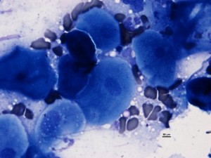
The Cytology
Squamous metaplasia with neutrophilic inflammation.
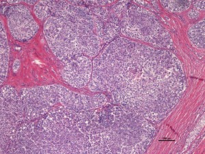
The Biopsy
The testicular neoplasm was composed of tubules of neoplastic cells, with robust fibrous tissue stroma. Diagnosis: Sertoli cell tumor (SCT)

Squamous metaplasia of the prostate gland associated with this type of tumor (SCT) is well-described and is caused by hormonal stimulation, typically excess estrogen, produced by the neoplastic cells.
Once the testicle and tumor were removed, the prostate shrunk and the hair grew back!
I did not see a skin biopsy in this case. But really, I didn’t need to. Others signs of feminization such as gynecomastia (enlarged mammary tissue) and occasionally bone marrow suppression due to the high estrogen levels can also be seen. The CBC changes vary with the duration of estrogen exposure by the bone marrow cells.
Inhibin is another hormone produced by sertoli cells, and can be measured in blood and via immunohistochemical stains, though not completely specific for this type of tumor.
What is new with the diagnosis of sertoli cell tumors?
Sertoli cells of fetal and immature dogs express Anti-Muellerian Hormone (AMH), a feature which is lost as the dog matures. In one study, all sertoli cell neoplasms expressed AMH via immunohistochemical staining of formalin fixed tissue, while control adult dog testicular tissue did not*.
In humans, serum levels of AMH are inversely related to testosterone levels. Likewise in dogs, the testosterone level is often low and the estrogen level high in those with SCT.
Differentiation of the three common canine testicular tumors, the other two being seminomas and interstitial cell tumors, is thankfully usually fairly straightforward based on morphology. The literature is somewhat conflicting in the molecular and hormonal tests used to differentiate these neoplasms. Seminomas in general express c-kit immunohistochemical staining at a higher rate than the other tumor types, while interstitial cell tumors show high serum level and immunohistochemical staining pattern of inhibin-alpha, which is also seen in sertoli cells**.
While the hormone levels were not measured in the dog in my case, the hormonal pattern of hair loss and squamous metaplasia support a state of hyperestrogenism.
Thankfully, resolution of the clinical signs with the return to a normal lush, furry body support normalization of the hormone levels.
Audrey Baldessari, DVM, Diplomate, ACVP (Anatomic and clinical)
*Journal of Comparative Pathology, Banco et al, 2012 Vol 46 (1) pages 18-23
**Journal Vet Science, Yu et al, March 2009 Vol 10(1) pages 1-7

